image of soil quality testing in a laboratory

Same tissue. More Insights.

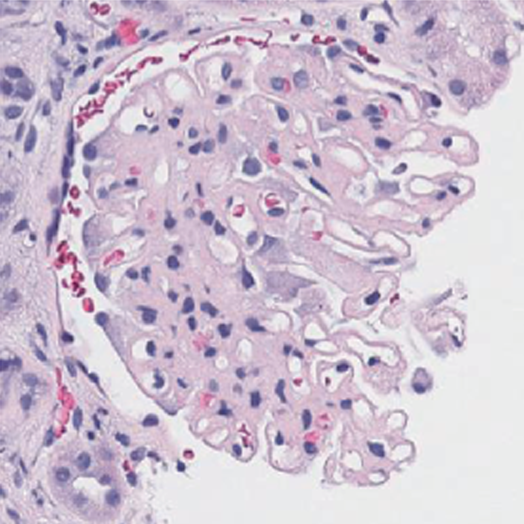
Pictor Labs' virtual staining technology generates additional histologic views (including H&E, special, IHC-like, and other stain representations) from the specimens and slides laboratories already have, with results available in minutes rather than days. Our models are trained on expertly curated, co-registered image pairs to provide visually comparable, consistent virtual stains that support diverse research and downstream workflows, without consuming additional tissue.
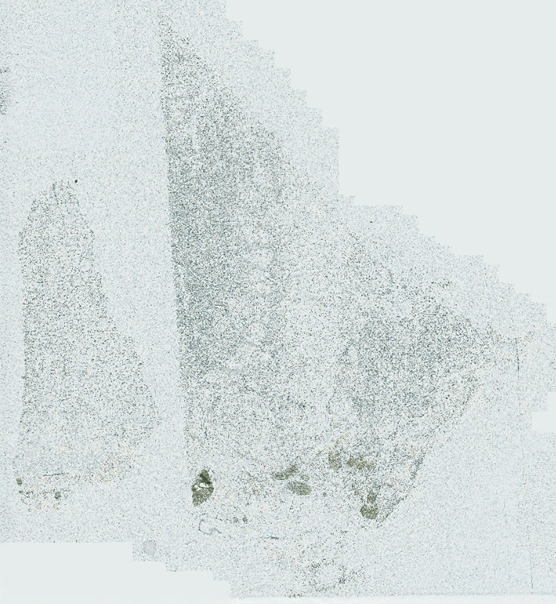
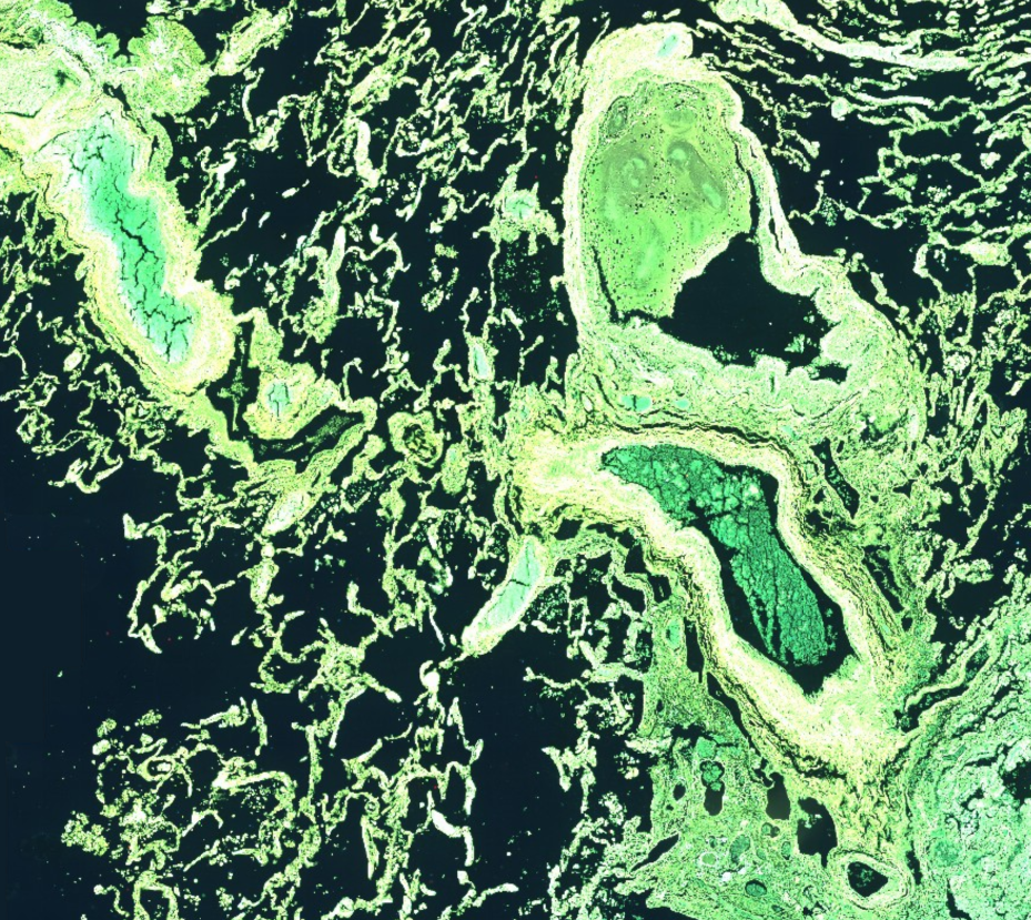
Virtual Staining Scroll Animation
Specimen Acquisition Slide Prep H&E Staining WSI Chemical Staining Pathologist Review Molecular Testing Report Virtual Staining Pathologist Review On Demand IHC & Special Staining

Scroll to see how virtual staining transforms the workflow

On-premises

Cloud

Best for

Full data control, controlled lab environments, or existing imaging pipelines.

Best for

Rapid scaling and collaborative, multi-site studies.

Key benefit

Fast, local processing with total data oversight.

Key benefit

Zero hardware burden; flexible, on-demand power.

Maintenance

Managed via your internal IT infrastructure.

Maintenance

Seamless updates without infrastructure overhead.

Built for scalable research and translational workflows

Pictor Labs replaces chemical staining with non-destructive virtual staining. Traditional staining consumes tissue with every iteration. Pictor Labs preserves tissue while expanding insight. Whether in a high-security lab or a distributed cloud environment, Pictor fits seamlessly into your workflow.

Each system is calibrated to match your lab’s diagnostic standard, ensuring pathologists see what they expect to see. Both deployment options deliver the same high-fidelity virtual staining outputs.

Ready to transform your lab’s throughput?

ClearStain

What You See Is What You Sequence

ClearStain™ generates a virtual H&E image directly from an unstained brightfield-scanned tissue section, preserving all material for downstream molecular analysis. Because the tissue is never chemically treated, everything available for extraction remains intact for NGS, spatial biology, and other ancillary studies. The virtual stain is generated from the exact section being used for sequencing, enabling precise annotation and tumor-rich area identification before macro-dissection. Tumor targeting and enrichment capabilities allow researchers and qualified reviewers to identify tumor-rich regions and exclude areas that would dilute sample purity, supporting more accurate downstream sequencing outcomes.

For Research Use Only.

Brightfield tissue image
Brightfield
Fibrosis
Inflammation
Tumor

DeepStain

One Slide Is All You Need

DeepStain™ generates multiple virtual stain outputs from a single unstained autofluorescence image, delivering multi-stained images from a single tissue section without chemical treatment or additional sectioning. A single unstained fluorescent scan serves as the input, from which DeepStain produces IHC-like and special stain outputs including virtual PanCK, Masson’s Trichrome, CD45, and others. Because no additional tissue is consumed in the process, the same section can support multiple stain representations, reducing the specimen burden of workflows that would otherwise require separate sections for each stain type.

For Research Use Only.

Pick an output stain, then hover or drag on the tissue to preview.

Autofluorescence tissue image
Autofluorescence
Tip: on touch devices, drag across the tissue to move the lens.

ReStain

Transform Chemical H&Es

ReStain performs virtual transformation of conventional H&E stained tissue into multiple special stain and IHC-like outputs from a single tissue section, without requiring additional slides or chemical processing. A ground truth H&E section serves as the input, from which ReStain generates virtual special stains and IHC-like markers including Trichrome, Reticulin, PanCK, CD45, and others across tissue types. For research programs that have already processed tissue to H&E, ReStain extends the analytical value of existing sections by enabling on-demand virtual restaining without consuming additional material.

For Research Use Only.

Pick an output stain, then hover or drag on the tissue to preview.

H&E tissue image
H&E
Tip: on touch devices, drag across the tissue to move the lens.

RetroStain

Transform Stains, Across Scanners and Modalities

RetroStain™ converts chemical or virtual stains across scanners and imaging modalities, without requiring additional tissue sectioning or processing. Color Correct normalizes whole slide images across brightfield scanner color profiles, enabling consistent visualization across multi-site or multi-instrument environments where different scanners produce different color renderings of the same tissue. IHC to H&E generates virtual H&E output from archival tissue where IHC is more readily available. mIF to IHC translates multiplex immunofluorescence results into IHC-like representations designed to support pathologist-friendly review, making mIF data more accessible for interpretation workflows that are oriented around conventional IHC appearance.

For Research Use Only.